Eye clinic Ladavac Pula Istra

DIAGNOSTICS AND TREATMENT OF EYE DISEASES
Conjunctivitis, keratitis, iridocyclitis, sty

Conjunctivitis

Conjuntivitis is an inflammation which includes intensification of tears, redness and the feeling of having sand in the eyes. It is mostly allergic, but it can also be caused by the dry eye syndrome or, more rarely, by a bacterial inflammation. It is treated depending on the cause.

Conjunctivitis

Keratitis

Keratitis is the inflammation of the transparent part of the eye called cornea. It is mostly caused by viruses and it can lead to vision opacity, intensification of tears and the feeling of having a foreign body in the eye. It is dangerous because it can return. Keratitis can sometimes appear with persons wearing contact lenses. It can be both bacterial and fungal and it can lead to an even permanent loss of vision.

Iridocyclitis

Iridocyclitis is the inflammation of the anterior part of the eye, the iris, and includes vision opacity, redness of the cornea and sometimes headache due to an increase in the eye pressure. It has to be treated for a longer period of time and it can cause vision opacity and an increase of the eye pressure. It can return periodically.

Sty

Sty (hordeolum) is an acute infection causing a red nodule at the edge of the eyelid.

Chalazion is a type of eye sty which became chronic. It is usually larger and it does not hurt. Sties and chalazions can be linked to blepharitis, a common problem causing inflammation of the eyelid. If it is not cured by eye drops, surgical removal and emptying of the sty are necessary.

Sty (hordeolum), Chalazion

DIAGNOSTICS AND TREATMENT OF EYE DISEASES
Eye pressure (glaucoma)

Glaucoma is a group of diseases when, due to high eye pressure, the head of the optic nerve and the visual field get damaged, which, if not treated, can lead to blindness.

Diagnosis, monitoring and glaucoma test

Glaucoma is easy to determine in the present times. The most reliable method is the OCT, a method most similar to a CT, but without radiation. The optic nerve is scanned and its degeneration monitored.

Measuring the visual field, eye pressure and eye angle are also ways of determining the presence of glaucoma. The complete examination at an glaucomatologist’s lasts half an hour and it will offer a certain answer to the query about the presence of glaucoma.

Glaucoma treatment

Glaucoma treatment is graded from medicines, through lasers to surgery. Sine glaucoma does not cause pain, people are often careless and think that they can control the situation with eye drops. However, by avoiding regular examinations where the doctor prescribes the eye drop dose to be applied, the patient can cause a serious damage because failure to comply with the doctor’s prescriptions is the most usual cause of blindness.

If the glaucoma eye drops are unpleasant or irritable, do not stop with the therapy without consulting your doctor about possible alternative solutions..

GLAUCOMA – the second most common cause of blindness in the world

Glaucoma is a type of eye disorder usually associated to a harmful increase of the interior eye pressure which can damage the optic nerve transporting the visual information to the brain. The problem occurs when the normal circulation of the aqueous humour between the eye chambers is impeded, and its accumulation raises the eye pressure. The high eye pressure gradually damages the optic nerve which leads to the loss of vision.

If glaucoma is not treated and controlled, the only thing expected in narrowing of the visual field.

The disease escalation can lead to blindness!

It is a very common disease, the second cause of blindness in the world. According to the World Health Organisation data, 4% of people in the world (65 million) suffers of glaucoma, and it has been estimated that by 2020 the glaucoma will be the cause of blindness for more than 11 million people around the world.

Glaucoma symptoms

Glaucoma is also called “the silent vision thief” because in most cases there is no pain or symptoms, until the loss of vision becomes obvious.

Due to this reason, glaucoma can usually not be noticed until the optic nerve becomes irretrievably damaged with a permanent loss of vision.

It sounds like a denouncing disease, but it does not have to be like that if detected on time. After reaching 40 years of age, people should do a more detailed examination which would determine if there is a glaucoma threat awaiting.

Contrary to chronic glaucoma, acute glaucoma had dramatic symptoms including vision opacity, diffusion of light, intensive pain. If you have similar symptoms, make an appointment at your eye doctor’s because if you do not react promptly, you could lose your vision in seven days!

DIAGNOSTICS AND TREATMENT OF EYE DISEASES
CHANGES CAUSED BY DIABETES

Diabetes

Diabetes is one of the most usual metabolic diseases characterised by higher values of sugar in the blood. If it is not treated, it leads to damages to smaller blood vessels in the kidneys, heart, extremities and eyes. In the eyes it causes bleeding in the posterior part of the eye, the retina, which may also lead to blindness. Diabetes is a dangerous disease because it gradually ruins one’s health, while patients are not aware of that due to the lack of pain or other signs which could warn about the advancement of the illness. It is difficult to heal because the key to successful treatment is great discipline in the patient’s behaviour, not only taking medicine (nutrition, life habits).

Diabetes and the eye

Diabetes can be the cause of cataracts on the eye, but also of other, more serious, changes on the posterior part of the eye, the retina. The eye blood vessels become narrower and more rigid due to diabetes, they easily break, and that leads to bleeding. Bleeding causes weakening of vision. It is treated with special medicine applied into the eye (anti VEGF therapy) and by laser, while the most serious cases are solved by special surgeries (vitrectomy). Occasional eye examinations are obligatory regardless of the patient’s good vision and overall condition because detecting eye diabetes (diabetic retinopathy) early significantly influences the results and prevents the loss of vision.

Diabetes can be the cause of cataracts on the eye

DIAGNOSTICS AND TREATMENT OF EYE DISEASES

Age-related macular degeneration (senile macular degeneration)

Senile macular degeneration is a disease which is significantly increasing due to longer lifespan. This is a disease occurring with older people regardless of their sex and causes the decline of central vision. People can see thanks to a small part in the centre of the eye, on the retina, called macula or macula lutea. This part of the eye consists of a large number of special cells which receive images and transport them to the brain. This part is void of blood vessels, but with age and due to genetic predisposition, senile changes start to occur in this area.

Senile macular degeneration is a disease which is significantly increasing due to longer lifespan
Patients come for an eye examination because they read with difficulty, they see intermittently and unevenly, while the advanced condition shows a central dark spot.

There are two subtypes of the disease. In the first case the central vision degenerates (atrophy) and it is called dry age-related macular degeneration. In the second case new blood vessels appear, they bleed and cause loss of vision (wet age-related macular degeneration). To set a diagnosis it is necessary to examine the posterior eye in mydriasis (pupil dilated), OCT of the macula and possibly the fluorescein angiography. The treatment is based on the diagnosis.

If degeneration of the cells occurs (wet form), polyvitamin therapy is applied, but when there is a macular bleeding, injections are applied which close the blood vessels and cause recession of bleeding (anti VEGF). Injections have to be applied each month during three months, and then treatment is continued based on the clinical image. Results depend on the pre-treatment findings and on reactions to medicine. Nevertheless, it should be emphasized that the patients have a weakened vision, but are not completely blind.

human eye parts - We see thanks to a small part in the centre of the eye, on the retina, called macula or macula lutea

DIAGNOSTICS AND TREATMENT OF EYE DISEASES

Vascular retinal diseases

The blood flow in the retina is very good, except for its central part. Like all areas where there is a thick network of blood vessels, they can also become occluded. Arteries (leading blood into the eye) and veins (leading blood out of the eye) are basic blood vessels.

Vascular retinal diseases
Occlusion (embolism) of the eye arteries

Occlusion of eye arteries is, in fact, an eye arrest. The whole area getting fed by the artery remains unfed and degenerates. It causes an abrupt and huge loss of vision or blindness. Arterial embolism occurs quickly and painlessly. There is not a right cure and a complete examination is necessary to avoid embolism on the other eye.

Occlusion of the eye veins (thrombosis)

Occlusion of the eye veins (thrombosis) is one of the most common vascular diseases of the eye. It also occurs suddenly and causes a more or less serious loss of vision. During thrombosis the blood cannot run out of the eye so it is accumulated in the retina which starts swelling. Usually it affects the vision centre with an edema. It is successfully treated with injections applied directly in the eye (anti VEGF or corticosteroids), but a long lasting therapy is needed. A systemic examination is also necessary to find the reason of thrombosis occurrence.

DIAGNOSTICS AND TREATMENT OF EYE DISEASES

Eye floaters

The eye is filled with a transparent liquid. This liquid has a regular structure, but by ageing this structure changes and floaters in the eye occur. Patients usually see them on a light background, but they do not cause weakening of vision. When floaters become thicker, appear suddenly and are followed by lightning, they can be a reason to worry about. Stronger opacity can be the sign of internal eye bleeding, inflammation and the possibility of formation of small holes on the retina. These holes are dangerous because they can cause the detachment of the retina or the occurrence of ablation. If timely recognised, laser treatment is used to protect the eye from the development of retinal ablation.

Macular membrane

Sometimes thickening of a delicate membrane can occur on the macula lutea. The membrane slowly shrinks and wrinkles the macula so patients see an uneven image and are not able to read clearly. The disease is diagnosed based on the clinical image and OCT of the macula. If it causes more serious disturbances, surgery can be also applied (“macular peeling”). Sometimes the macular membrane can spread so that a small hole appears (macular hole).

Ablation of the retina

When the internal layers of the eye detach from the external ones, it is called detachment or ablation of the retina. There are more types of ablation, but the most common one is the spontaneous ablation occurring without any clear reason. Holes on the peripheral part of the retina appear and they let liquids pass behind the retina and its slight detachment. The main symptoms are occurrence of floaters, lightning in front of the eye and vision opacity. As detachment becomes larger, the vision gets weaker and it feels like having “curtains in front of the eye.” This is a disease which could be cured by surgery.

Ablation of the retina

DIAGNOSTICS AND TREATMENT OF EYE DISEASES
DIAGNOSTICS AND TREATMENT OF VISION OPACITY CAUSED BY THE DEVELOPMENT OF CATARACT

The cataract is opacity of the lens causing vision opacity. This is the most usual eye disease and it requires surgical treatment. It develops gradually and it leads to painless vision opacity. In previous times practitioners waited the cataract to “ripen”, i.e. the patient to have a really bad vision, almost blindness, and then it was operated. Today these operations are routine. Good appliances and the ultrasound method give much better results and recuperation is quicker.

The operation technique comprises the removal of the opaque lens and putting an artificial lens into the eye. The surgery is conducted under local anaesthesia (eye drops), without injections. The eye is entered through a small hole and after cleaning the opaque lens, a flexible artificial lens of adequate dioptre is introduced, which means that after surgery patients do not need farsighted eyeglasses, or there is a minor dioptre solved by eyeglasses. This surgery is called facoemulsification (ultrasound surgery, popularly known as laser cataract surgery, which is a technically incorrect name).

Frequently asked questions:

Kada treba operirati mrenu?

Mrena uzrokuje zamućenje vida te se operira kada smeta, odnosno kada poremeti kvalitetu života. To nije tumorozna tvorba koja zahtijeva hitnoću, ali niti ne treba čekati predugo, jer nas slab vid ograničava u svakodnevnome životu ( izbjegavamo vožnju, slabije vidimo čitati, mutnije vidimo na daleko, imamo zablještenja)

When should cataract be operated?

Cataract causes vision opacity, so it has to be operated when it starts causing inconveniences or when it disrupts the quality of life. This is not an emergent cancerogenous formation, but still it should not take long to solve it, because poor vision is restrictive for our everyday life (we avoid driving, we can hardly read, far vision is blurry, we see flashings).

What are the complications?

Although it is a routine operation, it still is an operation, and each operation can be risky. Complications can be intraoperative (during surgery) and postoperative. The most usual complication is the failure to implant the lens. The patient cannot see and the operation has to be repeated (secondary implantation), but other complications can also occur (bleeding, inflammation, increase of eye pressure). Postoperative complications are longer periods of corneal opacity, wrong artificial lens dioptre, move of the artificial lens and inflammations. The operation is conducted in an operating theatre, under sterile conditions, because inflammation can cause complete blindness. We have to be aware of the fact that a cataract operation can lead to complete blindness (rarely).

What are the degraded conditions for the operation?

The principle is to open the small bag through a hole, with dilated pupils. The opaque lens is then pounded and aspirated by the ultrasound and an artificial lens is then put into the eye. If you have a ripe cataract (a hard one), high eye pressure, high dioptre, if your pupil does not dilate or you have a relaxed iris, the operation could be harder with higher incidence of complications.

How do I prepare for the operation?

First you have to understand that the cataract causes inconveniences, do not let anyone talk you into the operation. The operation is much easier if you decided to undergo surgery yourself. It is necessary to undergo preoperative examinations (glaucoma, macular degeneration), biometry has to be done (determining the exact dioptre of the lens to be implanted) to achieve the best possible vision without glasses.

On the operation day you will take your everyday therapy and have a light breakfast. In the doctor’s office patients usually get a tranquilizer and eye drops for the pupil dilation are applied.

After the operation you have to keep to the doctor’s advice which will speed up recovery and diminish the possibility of infections. That means that you will have to apply eye drops against infections and inflammations regularly, be at rest and avoid doing hard physical works. In case of any disturbances or doubts on complications, you have to contact your doctor without delay.

The period of recovery lasts from eight to 10 days and after that you can slowly return to your everyday rhythm. After two or three weeks, eyeglasses are ordered if necessary.

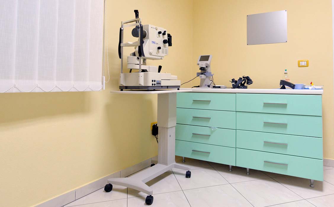
What kind of lenses are used in cataract surgery?

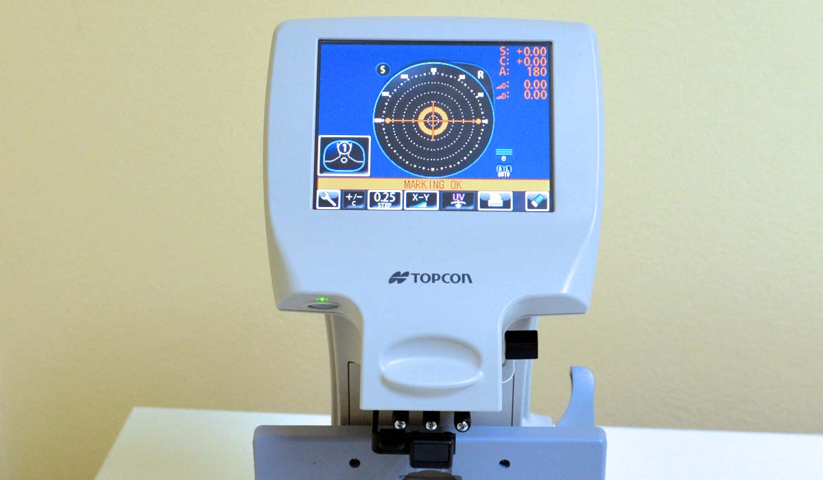
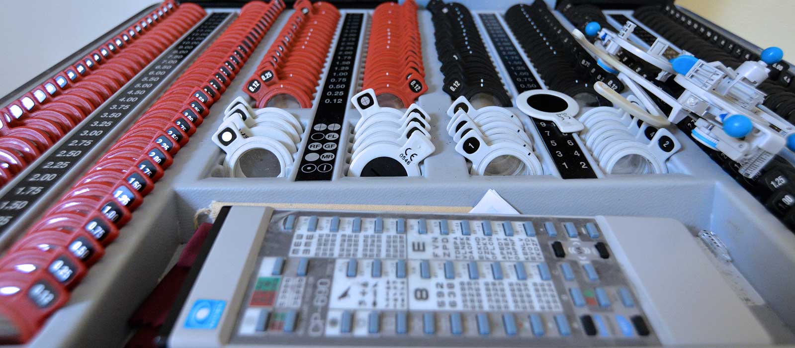
These are intraocular artificial lenses which means they are introduced in the eye, in the spot of the removed opaque lens and they are not extracted any more (they are not contact lenses). There are more types of lenses. The basic ones are those which give back the ability to see far objects, while eyeglasses (monofocal) are used for near vision. If the patient has higher cylinders, then lenses with cylinders (toric) are applied, and if patients do not want to wear glasses, then multifocal lenses are applied (with dioptre to be able to see far objects, to work on the computer and to see close objects). It is important to determine the intraocular lens dioptre. This is done by biometry, using a special appliance (each eye has its own dioptre, there are no standard dioptres).

Can cataract reappear?

No, it cannot, but the bag into which the artificial lens has been implanted can become turbid which is solved by laser cleaning of the posterior capsule of the bag turbidity. This is a simple and fast healing method, not an operation. The YAG Laser capsulotomy is done outpatiently.

Close